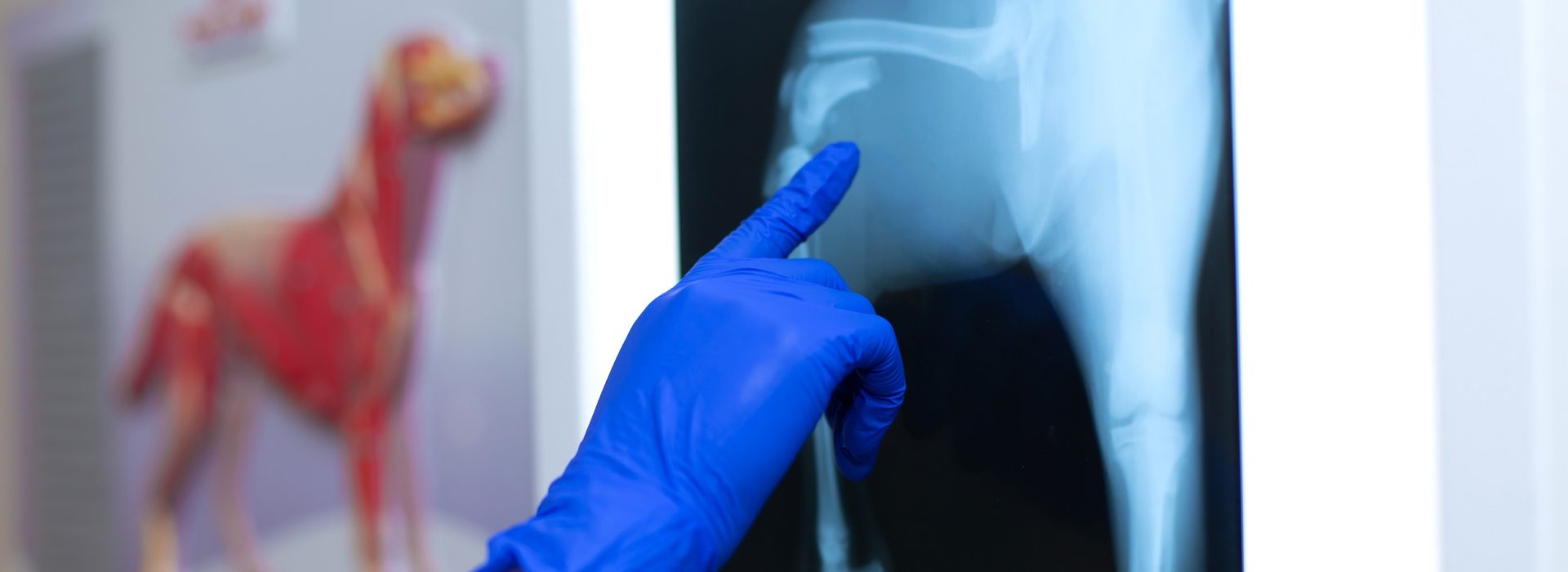
Ultrasounds and radiographs are great diagnostic tools in veterinary care. Radiographs are also referred to as X-rays. Both tools allow veterinarians to get information about your pet’s internal organs without surgery or any pain. Our veterinarians have received special training to interpret the images taken from radiographs and ultrasounds in order to properly diagnose your pet.
What can ultrasounds and X-rays diagnose in my pet?
Ultrasounds are often used to examine soft tissue. Ultrasounds can be used to detect:
- Tumours
- Pregnancies
- Fluids and cysts
X-rays can be used to identify:
- Arthritis
- Spinal cord disease
- Bladder stones
- Hip dysplasia
How do ultrasounds and X-rays work in my pet?
To perform an ultrasound a sonographer applies gel to a certain area on your pet. A handheld tool called a transducer is moved strategically over the gel. This transducer produces sound waves that echo and reveal what is at that site. The images are shown on a monitor that is connected to the transducer.
X-rays use a small amount of radiation to capture images as it passes through your pet. The amount of radiation is so small that it poses no risk to your pet even after multiple uses. The three most common types of X-rays are abdominal, chest and orthopedic.
Does my pet need to fast before getting an ultrasound or X-ray?
The only requirement for ultrasounds and X-rays is to shave the hair off a particular area. Removing the hair allows the images from both procedures to develop better. This is not something that you need to do on your own, as our veterinary care team will shave the area. If your pet is particularly anxious, or if they have to be placed in an awkward position for the scans, sedation may be a better option to prevent unnecessary stress for them. In this instance, fasting for this type of procedure will be required.


